PaNanoMRI

Precision imaging of premalignant cancer

Source: mrimaster

Current imaging tests provide inadequate sensitivity/specificity for detection of pre-malignant pancreatic cancer lesions because they are either too small (e.g. on MRI/CT) or isointense/isodense to normal tissue. This potentially leads to missed diagnoses on imaging in at-risk patients. This project gathers a team of clinical/non-clinical scientists to work on this challenge, more specifically, through the combination of two novel non-invasive approaches to increase the diagnostic yield of MRI in patients with early malignant disease: 1- Development of new multifunctional targeted nanoparticles for detection of pre-malignant pancreatic lesions, and 2- Improvement of MRI diagnostic yield through the application of super-resolution reconstruction (SRR) and quantitative MRI from MR Fingerprinting (MRF) for reproducible and rapid scanning.

Pancreatic cancer has the lowest survival of all common cancers, with five-year survival less than 7%, and the 5th biggest cancer killer in the UK [PCUK]. Early diagnosis is crucial to improve survival outcomes for people with pancreatic cancer; with one-year survival in those diagnosed at an early stage six times higher than one-year survival in those diagnosed at stage four. However, around 80% of patients are not diagnosed until the cancer is at an advanced stage. At this late stage, surgery/treatment is usually not possible. Not only do we need to have the tools and knowledge to diagnose people at an earlier stage, but we also need to make the diagnosis process faster so that we don’t waste any precious time in moving people onto potentially life-saving surgery or other treatments.

Collaborators

UCL Hospitals NHS Trust, universities of Strathclyde, Glasgow, Liverpool and Imperial College London.

Contact

Mohammad Golbabaee

Deep Compressive Quantitative MRI

Making MRI fast and quantitative

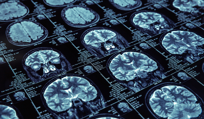
Closeup of X-ray photography of human brain

This interdisciplinary project focuses on the development of next-generation fast quantitative MRI precision imaging solutions based on AI and compressed sensing.

Magnetic resonance imaging (MRI) has transformed the way we look through the human body, noninvasively, making it the gold-standard imaging technique for diagnosis and monitoring of many diseases. However, conventional MRI scans do not produce “quantitative” i.e. standardised measurements, and therefore it is difficult to compare MRI images acquired at different hospitals, or at different points in time, limiting the potential of this imaging technology for advanced diagnostic and monitoring precision.

Quantitative MRI (qMRI) aims to overcome this problem by providing reproducible measurements that quantify tissue bio-properties, independent of the scanner and scanning times. This can transform the existing scanners from picture-taking machines to scientific measuring instruments, enabling objective comparisons across clinical sites, individuals and different time-points. Despite established benefits in precise evaluation of diseases (e.g., cancer, cardiac, liver, brain disorders), qMRIs suffer from excessively long scan times that currently obstruct their wide adoption in clinical routines. This project works with some of the world’s top organisations in healthcare research towards solving this challenge and enabling the qMRI scans to become substantially faster, more patient-friendly and more affordable and accessible.

Collaborators

GE Healthcare, UCL’s Centre for Medical Imaging, IRCCS Stella Maris-IMAGO7, University of Zurich.

Funding

£340k, EPSRC

Contact

Mohammad Golbabaee

Convolutional Sparse Coding based framework for the diagnosis of Alzheimer’s Disease


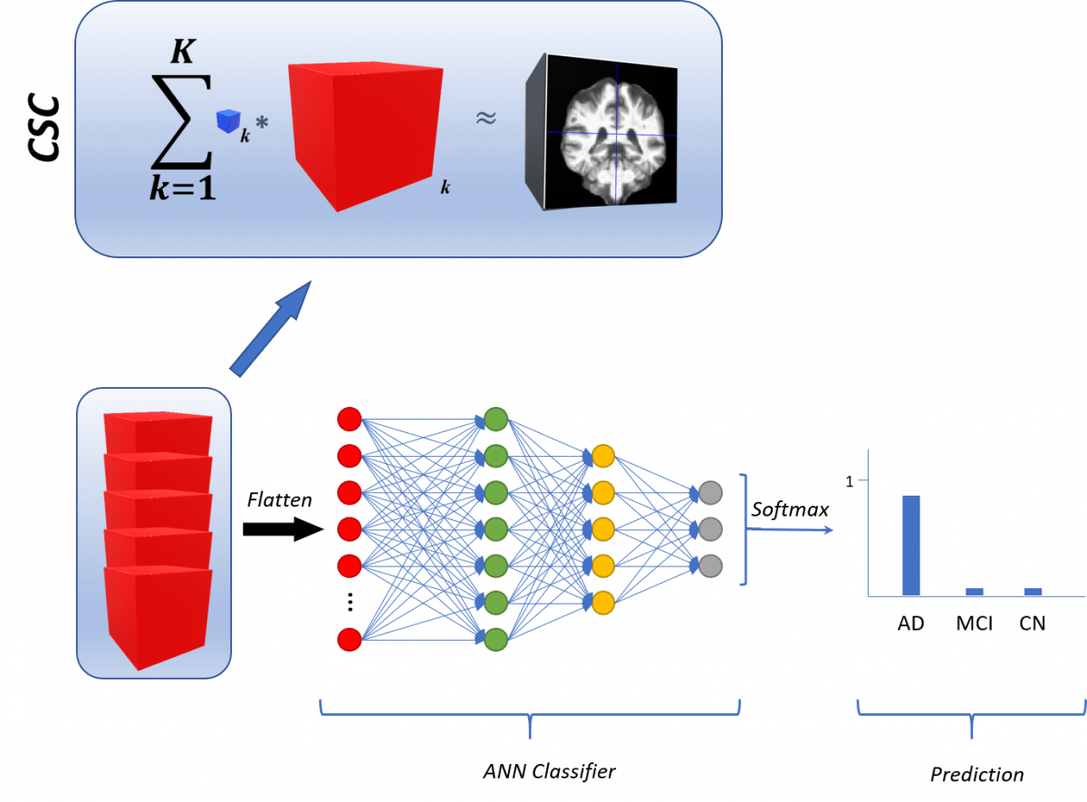
The learning and selection of features are leveraged to the Convolutional Sparse Coding approach, which is applied to the 3-dimensional MRIs in the dataset. They can be fed to any classifier, a fully connected neural network is shown here. The output corresponds to the prediction of the model for a given brain scan.

Diagnosis in medicine can be further aided by the automated inspection of structural images from the various body parts. Thus, the automated algorithm should be able to learn features from the images such that they will further aid in the accurate distinction of the disease, if present, from the image. This process can be done end-to-end using state-of-the-art techniques such as Convolutional Neural Networks (CNNs). In this work, we propose an alternative algorithm in which the diagnosis is performed as a two-step process. First, we learn the features from the 3-dimensional data, structural Magnetic Resonance Imaging (MRI) scans via Convolutional Sparse Coding (CSC). The features learned are then fed into some classifier –even CNNs—to fit a model able to differentiate the classes of interest, which correspond to the different diagnosis the patient could have for a particular disease

Contact

Perla Mayo, Alin Achim

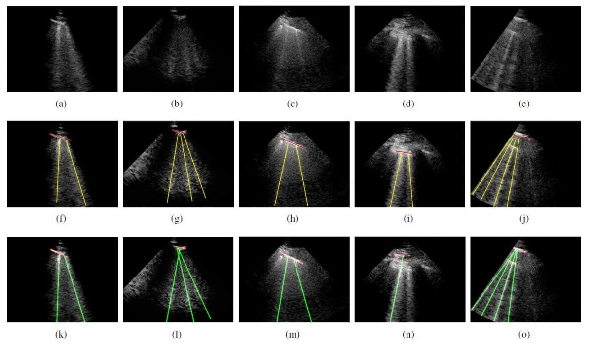
Line Artefacts Quantification for Lung Ultrasound Images via Non-convex Regularisation

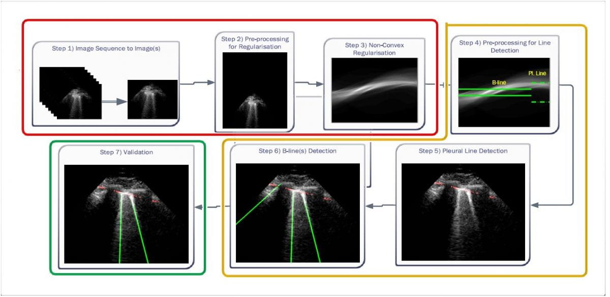
In this project, we present a novel method for line artefacts quantification in lung ultrasound (LUS) images of COVID-19 patients. We formulate this as a non-convex regularisation problem involving the heavy-tailed Cauchy-based penalty function, and the inverse Radon transform.

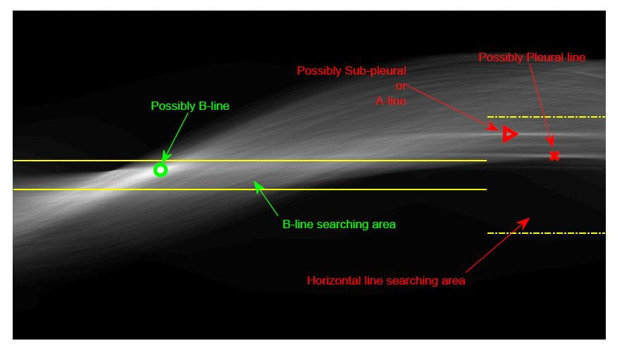
We employ a simple local maxima detection technique in the Radon transform domain, associated with known clinical definitions of line artefacts.

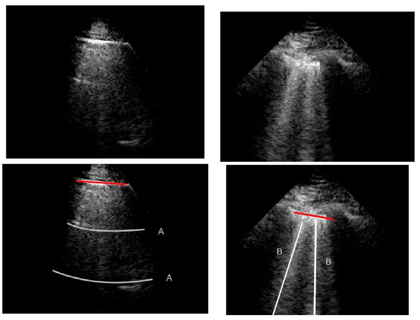
Line Artefacts in LUS Images
Line artefacts in Radon Domain

Despite being non-convex, the proposed technique is guaranteed to convergence through our proposed Cauchy proximal splitting (CPS) method, and accurately identifies both horizontal and vertical line artefacts in LUS images. In order to reduce the number of false and missed detection, our method includes a two-stage validation mechanism, which is performed in both Radon and image domains.

We evaluate the performance of the proposed method in comparison to the current state-of-the-art B-line identification method and show a considerable performance gain with 87% correctly detected B-lines in LUS images of nine COVID-19 patients.

Detection results. (a)-(e) Original Images. (f)-(j) Ground truth. (k)-(o) The proposed method.

Further information

[1] Karakuş, O., Anantrasirichai, N., Aguersif, A., Silva, S., Basarab, A., & Achim, A. (2020). Detection of Line Artifacts in Lung Ultrasound Images of COVID-19 Patients Via Nonconvex Regularization. IEEE Transactions on Ultrasonics, Ferroelectrics, and Frequency Control, 67(11), 2218-2229.

Talk

Talk by Dr Oktay Karakus in Acoustics Virtually Everywhere The 179th Meeting of the Acoustical Society of America 7-11 December 2020

https://player.vimeo.com/video/479873161

Contact

Oktay Karakuş, Pui Anantrasirichai, Alin Achim

Code

O Karakus, A Achim. (2020): QuantLUS – CPS v1.0 https://doi.org/10.5523/bris.z47pfkwqivfj2d0qhyq7v3u1i

Funding

This work was supported in part by
** the UK Engineering and Physical Sciences Research Council (EPSRC) under grant EP/R009260/1,
** an EPSRC Impact Acceleration Award (IAA) from the University of Bristol, ** a Leverhulme Trust Research Fellowship to Achim (INFHER).

Ship Wake Detection in SAR Images of the Sea Surface

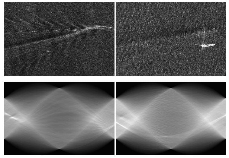
Example images including ship wakes and their Radon transform. (Upper Left) – Image includes all 5 ship wakes. (Upper Right) – Image does not include Kelvin wake. (Lower row) – Radon transforms of the images above. Both images are TerraSAR-X Stripmap products with HH polarisation and 3m by 3m resolution.

Ship wake detection is essential for extracting information on the wake generating vessels in order to analyse synthetic aperture radar (SAR) images of the sea surface. One possibility is to assume a linear model for wakes, in which case detection approaches are based on transforms such as Radon and Hough. These express the bright (dark) lines as peak (trough) points in the transform domain.

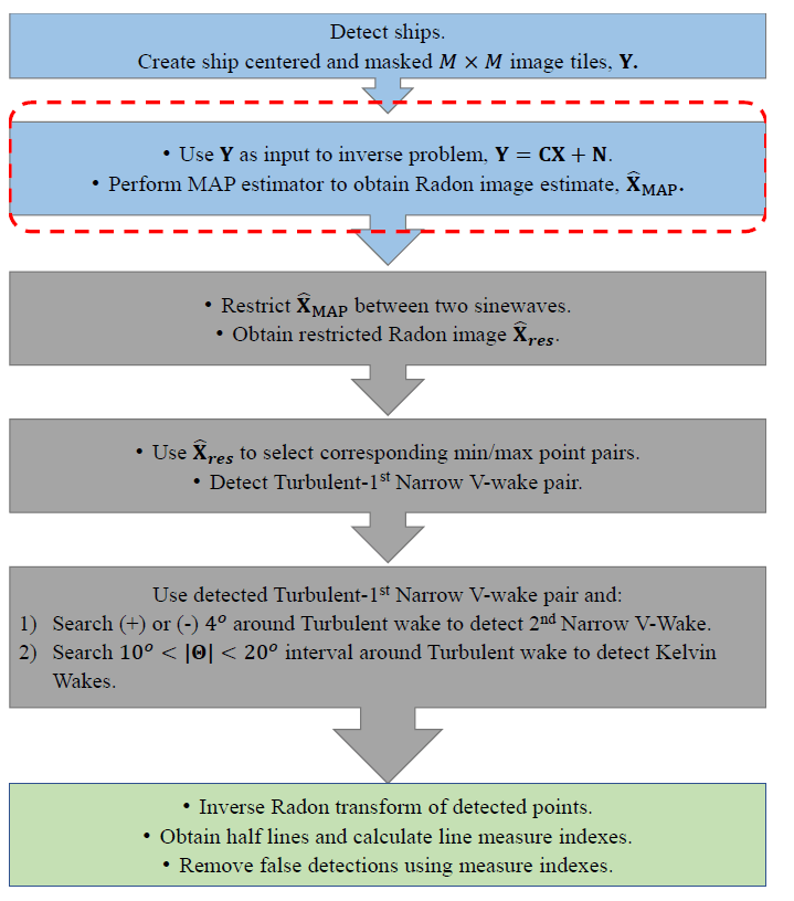
In this project, ship wake detection is posed as an inverse problem, which the associated cost function has taken various forms, such as

** Generalized minimax concave (GMC) function [1,2],
** Non-convex Cauchy-based penalty function [3,4],
** Hybrid penalty function which combines the Cauchy-based penalty with a 2-D DWT based term [5].

Despite having a non-convex regularizer, we provide mathematically consistent solutions, by either

** keeping the overall cost function to be convex [1,2], OR
** setting model parameters to guarantee the convergence [3-5].

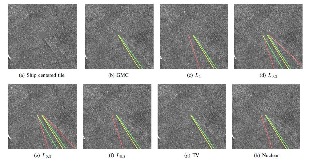
To quantify the performance of the proposed methods, various types of SAR images are used, corresponding to TerraSAR-X, COSMO-SkyMed, Sentinel-1, and ALOS2. The performance of various priors in solving the proposed inverse problem is first studied by investigating the GMC along with the L1, Lp, nuclear and total variation (TV) norms.

Algorithm

Example Results

Further information

[1] Karakuş, O., & Achim, A. (2019, May). Ship wake detection in X-band SAR images using sparse GMC regularization. In ICASSP 2019-2019 IEEE International Conference on Acoustics, Speech and Signal Processing (ICASSP) (pp. 2182-2186). IEEE.

[2] Karakuş, O., Rizaev, I., & Achim, A. (2019). Ship Wake Detection in SAR Images via Sparse RegularizationIEEE Transactions on Geoscience and Remote Sensing58(3), 1665-1677.

[3] Yang, T., Karakuş, O., & Achim, A. (2020, October). Detection of ship wakes in SAR imagery using Cauchy regularisation. In 2020 IEEE International Conference on Image Processing (ICIP) (pp. 3473-3477). IEEE.

[4] Karakuş, O., & Achim, A. (2020). On solving SAR imaging inverse problems using nonconvex regularization with a Cauchy-based penaltyIEEE Transactions on Geoscience and Remote Sensing.

[5] Ma, W., Achim, A., & Karakuş, O. (2020). Exploiting the Dual-Tree Complex Wavelet Transform for Ship Wake Detection in SAR ImageryarXiv preprint arXiv:2012.06663.

Contact

Oktay Karakuş, Tianqi Yang, Wanli Ma, Igor Rizaev, Alin Achim

Code

O Karakus, and A Achim. (2020): “AssenSAR Wake Detector.”
https://doi.org/10.5523/bris.f2q4t5pqlix62sv5ntvq51yjy

Funding

This work was supported by the UK Engineering and Physical Sciences Research Council (EPSRC) under grant EP/R009260/1 (AssenSAR).

CauchySAR: SAR Imaging Inverse Problems via Cauchy Proximal Splitting

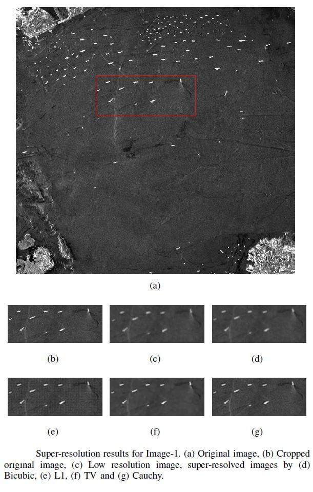
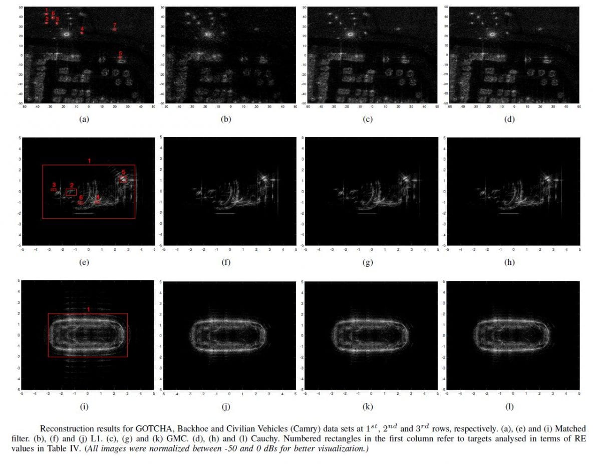
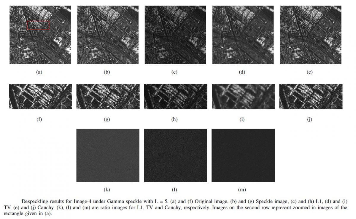
In this project, we investigate solutions to a number of inverse problems encountered in SAR imaging. We propose a convex proximal splitting method for the optimization of a cost function that includes a non-convex Cauchy-based penalty. The convergence of the overall cost function optimization is ensured through the careful selection of model parameters within a forward-backwards (FB) algorithm. The performance of the proposed penalty function is evaluated by solving three standard SAR imaging inverse problems, including

** Super-resolution,
** Image formation,
** Despeckling,
** Ship wake detection for maritime applications.

The proposed method is compared to several methods employing classical penalty functions such as total variation (TV) and L1 norms, and to the generalized minimax-concave (GMC) penalty. We show that the proposed Cauchy-based penalty function leads to better image reconstruction results when compared to the reference penalty functions for all SAR imaging inverse problems in this paper.

Super-resolution

Image Formation

De-speckling

Ship wake Detection

Further information

[1] Karakuş, O., & Achim, A. (2020). On solving SAR imaging inverse problems using nonconvex regularization with a Cauchy-based penaltyIEEE Transactions on Geoscience and Remote Sensing.

[2] O. Karakuş, I. Rizaev and A. Achim, “A Simulation Study to Evaluate the Performance of the Cauchy Proximal Operator in Despeckling SAR Images of the Sea Surface,” IGARSS 2020 – 2020 IEEE International Geoscience and Remote Sensing Symposium, Waikoloa, HI, USA, 2020, pp. 1568-1571, doi: 10.1109/IGARSS39084.2020.9323696.

Contact

Oktay Karakuş, Igor Rizaev, Alin Achim

Funding

This work was supported by the UK Engineering and Physical Sciences Research Council (EPSRC) under grant EP/R009260/1 (AssenSAR).