PaNanoMRI

Precision imaging of premalignant cancer

Source: mrimaster

Current imaging tests provide inadequate sensitivity/specificity for detection of pre-malignant pancreatic cancer lesions because they are either too small (e.g. on MRI/CT) or isointense/isodense to normal tissue. This potentially leads to missed diagnoses on imaging in at-risk patients. This project gathers a team of clinical/non-clinical scientists to work on this challenge, more specifically, through the combination of two novel non-invasive approaches to increase the diagnostic yield of MRI in patients with early malignant disease: 1- Development of new multifunctional targeted nanoparticles for detection of pre-malignant pancreatic lesions, and 2- Improvement of MRI diagnostic yield through the application of super-resolution reconstruction (SRR) and quantitative MRI from MR Fingerprinting (MRF) for reproducible and rapid scanning.

Pancreatic cancer has the lowest survival of all common cancers, with five-year survival less than 7%, and the 5th biggest cancer killer in the UK [PCUK]. Early diagnosis is crucial to improve survival outcomes for people with pancreatic cancer; with one-year survival in those diagnosed at an early stage six times higher than one-year survival in those diagnosed at stage four. However, around 80% of patients are not diagnosed until the cancer is at an advanced stage. At this late stage, surgery/treatment is usually not possible. Not only do we need to have the tools and knowledge to diagnose people at an earlier stage, but we also need to make the diagnosis process faster so that we don’t waste any precious time in moving people onto potentially life-saving surgery or other treatments.

Collaborators

UCL Hospitals NHS Trust, universities of Strathclyde, Glasgow, Liverpool and Imperial College London.

Contact

Mohammad Golbabaee

Deep Compressive Quantitative MRI

Making MRI fast and quantitative

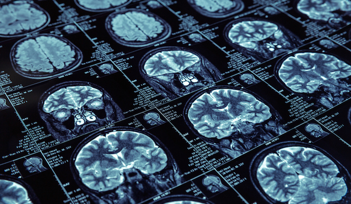
Closeup of X-ray photography of human brain

This interdisciplinary project focuses on the development of next-generation fast quantitative MRI precision imaging solutions based on AI and compressed sensing.

Magnetic resonance imaging (MRI) has transformed the way we look through the human body, noninvasively, making it the gold-standard imaging technique for diagnosis and monitoring of many diseases. However, conventional MRI scans do not produce “quantitative” i.e. standardised measurements, and therefore it is difficult to compare MRI images acquired at different hospitals, or at different points in time, limiting the potential of this imaging technology for advanced diagnostic and monitoring precision.

Quantitative MRI (qMRI) aims to overcome this problem by providing reproducible measurements that quantify tissue bio-properties, independent of the scanner and scanning times. This can transform the existing scanners from picture-taking machines to scientific measuring instruments, enabling objective comparisons across clinical sites, individuals and different time-points. Despite established benefits in precise evaluation of diseases (e.g., cancer, cardiac, liver, brain disorders), qMRIs suffer from excessively long scan times that currently obstruct their wide adoption in clinical routines. This project works with some of the world’s top organisations in healthcare research towards solving this challenge and enabling the qMRI scans to become substantially faster, more patient-friendly and more affordable and accessible.

Collaborators

GE Healthcare, UCL’s Centre for Medical Imaging, IRCCS Stella Maris-IMAGO7, University of Zurich.

Funding

£340k, EPSRC

Contact

Mohammad Golbabaee